When your pet feels unwell but cannot tell you what hurts, veterinarians need reliable ways to see inside their bodies. Diagnostic imaging for pets provides this crucial window into your companion’s internal health. These advanced technologies reveal problems invisible to physical examination alone.
Modern veterinary medicine has transformed how we detect and diagnose animal illnesses. What once required exploratory surgery now takes minutes with sophisticated imaging equipment. Your pet benefits from faster diagnoses and less invasive procedures.
This guide explores various imaging technologies, their applications, and how they help veterinarians identify health problems affecting your beloved companion quickly and accurately.
What Is Veterinary Diagnostic Imaging?
Diagnostic imaging encompasses technologies that create visual representations of your pet’s internal structures. These non-invasive or minimally invasive techniques allow veterinarians to examine organs, bones, and tissues without surgery.
Different imaging modalities serve specific purposes. Some excel at visualizing bones, while others reveal soft tissue abnormalities. Veterinarians select appropriate techniques based on suspected conditions.
These technologies have revolutionized veterinary care by enabling:
- Early disease detection
- Accurate diagnosis confirmation
- Treatment planning and monitoring
- Surgical preparation
- Cancer staging and evaluation
Common Types of Pet Imaging Technologies:
X-Ray (Radiography)
X-rays remain the most frequently used imaging tool in veterinary practice. This technology excels at visualizing bones, detecting fractures, and identifying foreign objects.
Radiographs also reveal:
- Heart size and shape abnormalities
- Lung conditions and fluid accumulation
- Bladder stones and kidney issues
- Intestinal blockages
- Arthritis and bone tumors
The procedure takes only minutes and typically requires no sedation for calm pets. Results appear immediately, allowing quick diagnostic decisions.
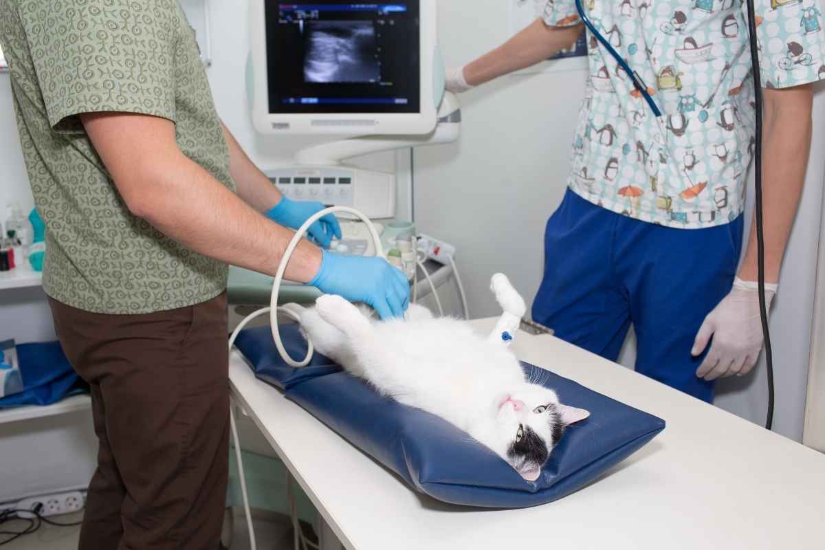
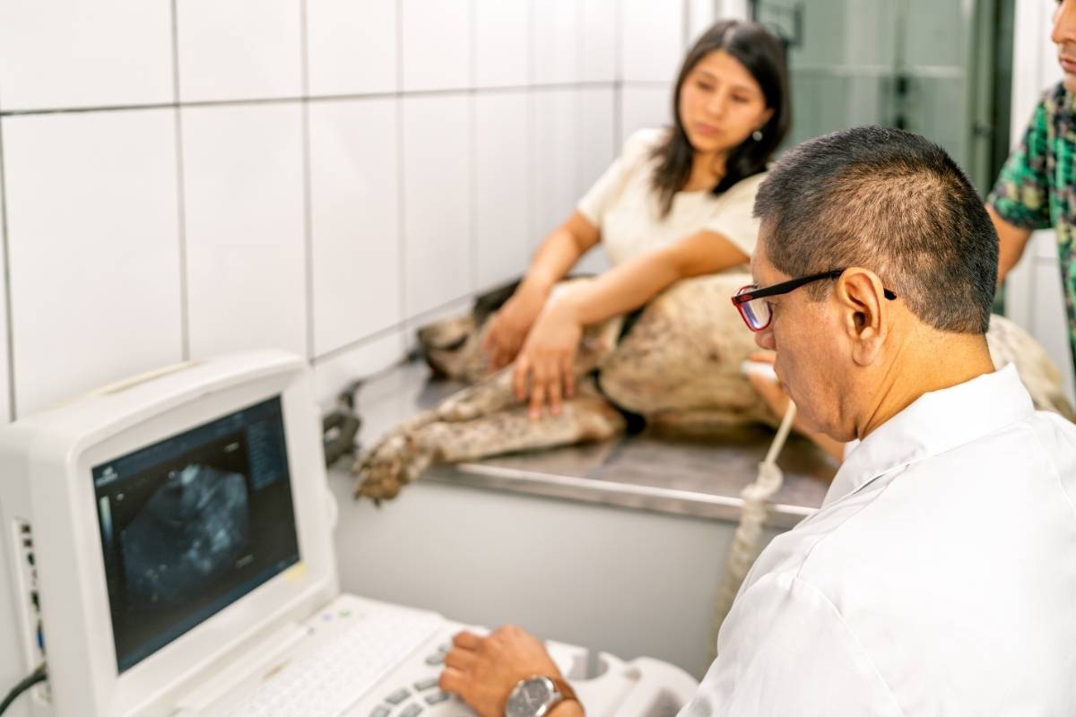
Ultrasound Imaging
Ultrasound uses sound waves to create real-time images of soft tissues and organs. This radiation-free technology proves invaluable for examining abdominal structures.
Veterinarians commonly use ultrasound for:
- Pregnancy confirmation and monitoring
- Liver and kidney evaluation
- Spleen and bladder assessment
- Heart function analysis (echocardiography)
- Tumor identification and biopsy guidance
Unlike x-rays, ultrasound shows organ movement and blood flow. This dynamic visualization helps diagnose conditions affecting organ function.
Advanced Imaging: CT and MRI
Computed tomography (CT) scans combine multiple X-ray images to create detailed cross-sectional views. This technology excels at complex bone evaluations and cancer staging.
Magnetic resonance imaging (MRI) provides exceptional soft tissue detail. Neurological conditions, spinal problems, and joint abnormalities become clearly visible through MRI technology.
These advanced modalities require anesthesia due to the need for complete stillness. However, they provide diagnostic information unavailable through other methods.
For comprehensive imaging services, the experienced team at Ark Veterinary Hospital & Urgent Care utilizes advanced technologies to diagnose your pet’s conditions accurately.
Conditions Commonly Diagnosed Through Imaging:
Orthopedic Problems
Imaging plays a crucial role in diagnosing bone and joint conditions. Fractures, dislocations, and developmental abnormalities appear clearly on radiographs.
Hip dysplasia evaluation requires specific X-ray positioning. Early detection allows intervention before severe arthritis develops.
Ligament tears and cartilage damage often require an ultrasound or an MRI for accurate diagnosis. These soft tissue injuries escape detection on standard X-rays.
Internal Organ Diseases
Organ enlargement, masses, and structural changes become visible through imaging. Veterinarians can assess liver disease, kidney problems, and splenic abnormalities without invasive procedures.
Cardiac imaging reveals heart enlargement, valve problems, and fluid accumulation. Early heart disease detection significantly improves treatment outcomes.
Cancer Detection and Staging
Imaging proves essential for cancer diagnosis and treatment planning. Veterinarians locate tumors, assess their size, and determine whether cancer has spread.
Chest radiographs check for lung metastasis. An abdominal ultrasound evaluates organ involvement. This information guides treatment decisions and prognosis discussions.
Trusted professionals specializing in veterinary diagnostic henrietta services can help identify concerning changes requiring immediate attention.
Preparing Your Pet for Imaging Procedures
Proper preparation ensures accurate results and reduces stress for your pet. Follow these guidelines:
Fasting Requirements: Many procedures require withholding food for 8-12 hours. Empty stomachs improve abdominal image quality.
Medication Considerations: Inform your veterinarian about all medications your pet takes. Some may require temporary discontinuation.
Sedation Preparation: If sedation is needed, follow pre-anesthetic instructions carefully. This typically includes fasting and limiting water.
Stay Calm: Pets sense owner anxiety. Your relaxed demeanor helps keep your companion comfortable during the appointment.
Understanding Your Pet’s Imaging Results
Veterinarians interpret imaging studies and explain findings in understandable terms. Don’t hesitate to ask questions about your pet’s results.
Request copies of images for your records. These become valuable references for future comparisons or specialist consultations.
Remember that imaging provides information, not always definitive diagnoses. Additional tests may be necessary to confirm suspected conditions.
Frequently Asked Questions:
Q1: Is diagnostic imaging safe for my pet?
Ans: Modern imaging technologies are very safe for pets. Radiation exposure from x-rays is minimal, and ultrasound uses no radiation. Your veterinarian takes appropriate precautions for all procedures.
Q2: Does my pet need anesthesia for imaging?
Ans: Most x-rays and ultrasounds require no anesthesia for cooperative pets. However, CT scans, MRIs, and some complex studies require sedation to ensure stillness and accurate images.
Q3: How long do imaging procedures take?
Ans: Simple X-rays take only minutes. Ultrasound examinations typically last 20-30 minutes. Advanced imaging like CT or MRI may require 1-2 hours, including anesthesia time.
Q4: Can imaging detect all health problems?
Ans: While imaging reveals many conditions, some problems require additional testing. Blood work, biopsies, and other diagnostics often complement imaging findings for a complete diagnosis.
Q5: How much does veterinary imaging cost?
Ans: Costs vary based on imaging type, body area examined, and geographic location. X-rays are generally most affordable, while an MRI represents a higher investment. Discuss pricing with your veterinary team beforehand.
Q6: When should my pet have diagnostic imaging?
Ans: Your veterinarian recommends imaging based on symptoms, physical examination findings, and medical history. Unexplained pain, breathing difficulties, abdominal swelling, or lameness often warrant imaging evaluation.
Related Articles